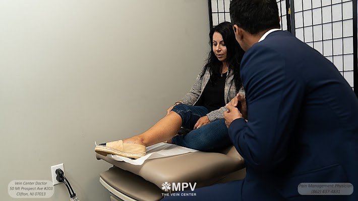
A single leg can hide three different problems at once, each demanding a different tool. That is the quiet reason many patients walk out of a vein therapy clinic with a plan that blends procedures rather than one silver bullet.
The real structure of vein disease, not just surface lines
What we see on the skin rarely tells the full story. Those purple spider webs on the calf can come from pressure higher up in the leg, often from reflux in the great saphenous vein or small saphenous vein. Reflux means valves that should direct blood upward have loosened, so blood drifts back toward the foot. That backflow raises pressure in the branches and tiny surface veins, creating varicosities and telangiectasias. This is venous hypertension, and over time it thickens vein walls, inflames the tissues, and can cause skin changes, swelling, or ulcers near the ankle.
A venous specialist doctor maps this network with duplex ultrasound. The scan studies direction and speed of blood, the diameter of veins, and whether branches connect to deep veins. Ultrasound also checks for clots. This map is the blueprint that guides a combination approach. If you only treat the visible veins without fixing the source of reflux, the symptoms usually creep back within months.
Why a single procedure often falls short
Every technique has strengths and blind spots. Endovenous laser or radiofrequency heat closes straight, incompetent trunks well, but it does not remove every bulging surface branch. Foam sclerotherapy reaches winding tributaries that a laser fiber cannot safely track, but foam alone may not handle a 10 millimeter saphenous trunk with heavy flow. Microphlebectomy plucks out ropey varicosities through pinholes, yet it cannot correct the underlying valve failure. Nonthermal adhesives avoid tumescent anesthesia, but they are limited by vein size and anatomy.


A vein therapy doctor balances these trade offs. The aim is not to stack procedures for the sake of volume. The aim is to match each step to the anatomy and clinical goals, then use the fewest steps that accomplish durable relief.
A practical framework for combination care
I find it helps to picture venous disease in layers: a source, a pathway, and a symptom layer.
- The source is usually truncal reflux in the great or small saphenous vein, or incompetent perforator veins feeding pressure into the skin. The pathway includes tortuous tributaries and clusters that carry the excess volume. They are often too winding for a straight catheter. The symptom layer is what you see and feel, from bulging cords and aching to spider veins and brown ankle skin.
Treat the source first so the pathway decompresses. Then address the remaining pathway segments and the cosmetic layer. That sequence cuts the number of sessions, shrinks anesthesia time, reduces bruising, and improves insurance approval when medical symptoms drive the case.
Common combinations and what each accomplishes
Here are patterns I use in a vein treatment center when the ultrasound map calls for more than one tool:
- Truncal ablation plus microphlebectomy. Endovenous laser or radiofrequency closes the refluxing saphenous trunk. In the same session or staged, an ambulatory phlebectomy doctor removes bulging tributaries through 2 to 3 millimeter incisions. This combination tackles both the source and the biggest symptomatic branches. It is reliable for CEAP C2 to C4 disease with large varicosities. Truncal ablation plus ultrasound guided foam sclerotherapy. When branches are too winding or too small for phlebectomy, a foam sclerotherapy doctor injects sclerosant under ultrasound guidance. This works well when there are clusters behind the knee or along the inner thigh that are awkward to remove through micro-incisions. Nonthermal glue or mechanochemical ablation plus foam. In patients who cannot receive tumescent anesthesia or who have very superficial trunks at risk for heat injury to the skin or nerves, a vein closure specialist may use cyanoacrylate adhesive or a rotating wire with liquid sclerosant. Residual tributaries often need a foam touch up. Perforator treatment plus wound care for ulcers. A venous ulcer doctor will often combine perforator closure with compression, debridement, and topical care. Once the ulcer begins to granulate, additional foam to nearby varices helps lower local pressure and speed healing. In a stubborn CEAP C6 case, staged care is safer than doing everything at once. Spider vein sclerotherapy after source control. A spider vein clinic should avoid injecting cosmetic webs until deeper reflux is under control. If done too early, the results fade. A vein injection specialist typically treats residual telangiectasias 6 to 12 weeks after the trunk work has settled.
These are not rigid recipes. A vascular and vein clinic will vary the mix based on vein diameter, depth from skin, tortuosity, prior surgery, and your goals.
Sequencing, staging, and why timing matters
Most venous care specialists prefer to start with the refluxing trunk. Closing that high flow pipeline reduces pressure in downstream veins within days. After two to four weeks, the leg often looks and feels different, and some branches soften or flatten enough to avoid additional work. When varices remain thick, a microphlebectomy specialist can remove them through vein specialist near me tiny nicks. Foam sclerotherapy may follow weeks later to polish what is left, including reticular feeders to spider veins.
Sometimes we stage even the trunk work. If both legs have long disease and you have a job that keeps you on your feet, doing one leg at a time eases recovery. In ulcer care, we treat the most hemodynamically significant segment first, add strict compression, and reassess the wound bed before the next step.
Timing also affects safety. Performing heavy foam injections immediately after truncal ablation can push sclerosant into freshly closed segments and raise the risk of pigmentation or matting. Spreading sessions by a few weeks lets the limb stabilize, reduces medication totals, and gives clearer targets on ultrasound.
Choosing techniques based on anatomy and life factors
A vein closure doctor has to weigh technical limits with patient realities. Here are examples I encounter at an outpatient vein clinic:
- Very superficial great saphenous vein within 5 millimeters of skin. Heat based closure risks a skin burn or nerve irritation. I lean to cyanoacrylate adhesive or mechanochemical ablation, then foam for branches. Severe tortuosity in the trunk. A laser fiber may not pass safely. I consider staged foam with careful dosing, or a hybrid with short segment ablation in straight sections and foam elsewhere. Tributaries clustered behind the knee. The small saphenous region hosts the sural nerve. Phlebectomy can irritate it. In many of these cases, ultrasound guided foam sclerotherapy offers a cleaner path. Previous stripping with recurrent reflux from groin neovascularization. Traditional vein stripping is less common now, but recurrences occur. Careful ultrasound mapping with a vein imaging doctor identifies aberrant channels. I often mix targeted foam with limited phlebectomy. Glue may help in short segments if accessible. Coexisting lymphedema or lipedema. These conditions complicate healing and swelling. I still address reflux, but I stage work conservatively, involve compression therapists, and plan for slower cosmetic improvement.
The role of compression and activity - still part of the mix
Even with modern techniques, a vein management specialist will often prescribe compression stockings in the early recovery phase. Graduated compression, usually 20 to 30 mm Hg, reduces bruising, speeds resorption of treated segments, and limits trapped blood that can cause tenderness. Walking the same day keeps blood moving in healthy channels. These are simple pieces of a combination plan, but they help as much as any catheter.
Patients sometimes ask if compression alone can fix varicose veins. It can control symptoms, which matters, but it does not reverse reflux. A combined plan might include a short course of compression while the leg calms after ablation, then selective injections or phlebectomy. The mix shifts as the leg changes.
Safety considerations when combining procedures
The safety record of minimally invasive vein treatments is strong. Complication rates for endovenous ablation sit in the low single digits. When combining steps, a vein procedure doctor watches total local anesthetic doses, total sclerosant volumes, and proximity to nerves and skin. Tumescent anesthesia has a lidocaine ceiling based on weight. Spreading sessions prevents cumulative overdosing.
Nerve irritation is uncommon but possible near the ankle with great saphenous work, and along the calf with small saphenous treatment. A careful venous surgeon uses ultrasound to track the nerve’s usual course and adjusts power settings and catheter depth. Thrombotic events are rare, particularly when the vein closure ends at least 2 centimeters below the deep system junctions. A deep vein thrombosis specialist would only add anticoagulation in narrow scenarios, such as a history of clotting, a known thrombophilia, or post procedure extension where surveillance scans dictate therapy.
Pigmentation, matting, and trapped blood after foam or liquid sclerotherapy respond to timely follow up. A vein injection doctor can evacuate trapped blood with a needle in clinic. Early intervention makes a difference in cosmetic outcomes.
Real cases that show why mixing tools helps
Case 1: A 46 year old teacher with aching, heaviness, and visible veins along the inner thigh and calf. Duplex shows great saphenous reflux from the groin to mid calf, with large tributaries feeding visible varices. We close the trunk with radiofrequency in one visit. Two weeks later, her symptoms improve and calf branches have softened, but two ropes remain tender. An ambulatory phlebectomy doctor removes them through eight pinholes. She returns 8 weeks later for limited liquid sclerotherapy to residual spider clusters around the knee. The three part plan totals under 90 minutes of procedure time, split across visits, and her insurance covers the first two steps based on symptoms and reflux documentation.
Case 2: A 63 year old man with a healed ankle ulcer and brown staining, CEAP C5. Ultrasound finds an incompetent perforator near the ulcer bed and reflux in the small saphenous vein. Because his small saphenous is very close to the skin at mid calf and he has diabetes with neuropathy, a vein closure specialist uses adhesive for the trunk and staged foam to clusters just above the ulcer. Compression and wound checks fill the intervals. The ulcer edges firm up after the perforator is addressed. A month later, he returns to work without bandages.
Case 3: A 35 year old runner with clusters behind the knee and tiny spider veins along the shin. Symptoms flare after long runs. The small saphenous vein shows segmental reflux and marked tortuosity that a laser catheter would struggle to navigate. An ultrasound guided sclerotherapy specialist performs two low volume foam sessions spaced three weeks apart. The runner wears knee high 20 to 30 mm Hg stockings for two weeks after each session and reduces hill workouts for a month. A third visit handles the spider veins with liquid sclerosant once deeper pressure has fallen. No thermal injury risk, and the nerve behind the knee is spared.
Case 4: A 58 year old woman with prior stripping 20 years ago and recurrent bulging veins. Mapping shows neovascular channels near the groin and reflux down a residual trunk remnant. A vein laser doctor cannot safely pass a fiber through the neovascular tangle. The plan mixes targeted foam to the aberrant clusters, short segment ablation lower in the thigh where the vein is straight, and later microphlebectomy of a lateral thigh bundle. It takes judgment and patience, but mixing techniques gives her relief without a large incision.
Cosmetic goals still benefit from medical sequencing
At a cosmetic vein specialist’s practice, patients often come in for spider veins. They are disappointed when we start the visit with ultrasound rather than a syringe. There is a reason. If a vein and circulation specialist finds reflux feeding the cosmetic network, treating the source first gives better, longer lasting cosmetic results. It also reduces the sclerosant volume needed on the skin layer, which lowers the risk of staining. When the deeper system is healthy, then a vein injection specialist can schedule two to four sessions for spider webs, spaced several weeks apart.
Insurance, documentation, and why the map matters
Payers look for medical necessity when approving ablation or phlebectomy. A vein diagnostic doctor’s ultrasound report is central. It should document reflux times, diameters, and affected segments in both the longitudinal and transverse planes. Symptom notes matter too, including heaviness, pain, edema, skin changes, and activity limits. Many plans require a trial of compression for a set interval, often 6 to 12 weeks, before authorizing ablation. Once approved, insurers usually expect treatment of the source first. Follow up branches may or may not be covered, depending on impact on symptoms. A good vein care provider will explain what is medical and what is cosmetic, then help you plan staging that makes sense clinically and financially.
What to ask a vein consultant doctor before saying yes
- Which vein is the main source of my reflux, and how will you treat it first What additional branches or spider veins might still need attention, and by which method How many sessions do you expect for my case, and in what sequence What are the risks for my anatomy, and how do you lower them What part is likely covered by insurance and what is optional or cosmetic
Where the setting matters
Combination care works best in a facility that can pivot between tools. A dedicated vein care clinic or vein health center has ultrasound in house and practitioners comfortable with ablation, injections, and phlebectomy. An outpatient vein specialist can manage most cases without general anesthesia, which keeps recovery short. For advanced disease, such as ulcers or recurrent clots, a vascular medicine specialist for veins or a venous surgeon may coordinate with wound care teams and, when needed, hematology. The setting does not need to be a hospital, but it should have protocols for rare complications and access to follow up scans.
When you visit a vein medical clinic, pay attention to how the team explains your map. A strong vein consultation specialist will use the scan to show why they recommend, for example, a small saphenous ablation, then a limited phlebectomy, then a spider vein touch up. If you only hear one procedure pitched to every patient, keep asking questions.
The technical side of matching tools
Here is how the common tools complement each other in daily practice:
- Endovenous thermal ablation. Laser or radiofrequency closes straight trunks reliably with high closure rates, typically above 90 percent at one year. Advantages include predictable outcomes and quick recovery. Limits include heat related risks near the skin or nerves, and difficulty in very tortuous segments. Nonthermal non tumescent closure. Cyanoacrylate adhesive or mechanochemical ablation avoids tumescent anesthesia and heat. Useful in superficial or nerve adjacent segments. Limits include vein size restrictions and cost considerations. Some insurers have narrower coverage for adhesives. Ultrasound guided foam sclerotherapy. Foam occupies more lumen than liquid and can be tracked in real time. Ideal for winding tributaries, perforators, and recurrent neovascular channels. Risks include pigmentation, matting, and rare clot extension. Dose control and spacing lower those risks. Microphlebectomy. Ambulatory phlebectomy removes bulky varices through micro incisions. It gives immediate contour change and relief of tenderness from tense varices. It pairs well with trunk closure. Bruising is common but fades in weeks. It is not a substitute for fixing reflux higher up.
A vein restoration specialist will often pick two of these for a single leg, applied in the right order. Add compression, walking, and follow up ultrasound, and you have a complete plan rather than a single shot.
Edge scenarios that demand tailored mixes
- Athletes and heavy laborers. Timing around seasons or work schedules matters. Combine treatments in fewer, longer sessions if safe, to minimize days off. A vein intervention specialist will still avoid overloading sclerosant dosing in a single day. Older patients with anticoagulation. A vein thrombosis doctor coordinates peri procedural management. Thermal ablation can proceed safely in many on blood thinners, but phlebectomy may bruise more. Staging lets the team adjust medications cautiously. Superficial vein thrombosis. If a superficial clot has formed in a varix, a superficial vein thrombosis doctor treats the acute phase first. Once inflammation resolves, closure of the source and removal or foam of the clot prone cluster helps prevent recurrence. Pigmentation prone skin. Lower sclerosant concentrations, careful avoidance of extravasation, and slower staging reduce staining risk. Cosmetic steps move to the end, once the leg is hemodynamically quiet.
Measuring success beyond a single before and after
Success is not just a photo of a smoother calf. It is heavier things becoming light again at 4 pm. It is walking upstairs without that deep ache. In clinic, we track symptom scores, calf circumference, healing of dermatitis or ulcers, and closure rates on ultrasound. Durable results correlate with correcting the source reflux first, then addressing what remains with the right adjunct. A vein care surgeon who treats only what the eye sees risks quick relapse.
Follow up matters. A vein screening specialist may scan at one week to rule out junction extension, at three months to confirm durable closure, and later if symptoms recur. If a new cluster appears in a different area, it is better to catch and treat it while small rather than wait for another full network to build.
How to choose a team for combined care
Look for a vein specialty clinic that:
- Performs its own duplex mapping and explains it in plain language Offers multiple modalities, not just one Schedules logical staging that treats the source before the surface Tracks outcomes with standardized measures and follow up scans Coordinates with wound care or thrombosis experts when needed
Titles vary. You may see a vein health doctor, a venous care physician, or a vascular vein surgeon. What matters is their breadth across techniques and their judgment in using them sparingly yet effectively.
The bottom line for patients
If your plan from a vein solutions clinic includes more than one step, it is usually a sign of careful thinking, not overtreatment. Vein disease rarely lives in a single segment. A venous reflux doctor uses ultrasound to find the source, then selects the least number of tools to fix the layers involved. Sometimes that is ablation alone. Often it is ablation plus a small adjunct, or staged foam for stubborn clusters. For spider veins, it may be a pause after source control, then a few quick sessions with a vein injection doctor.
Ask to see your map. Ask which step treats the cause and which tidies the effect. When the answers line up with your anatomy and symptoms, combination therapy stops feeling like more, and starts feeling like the right amount.